

A cholesteatoma is a collection of keratinized skin cells and debris that becomes trapped in the middle ear, mastoid, or nearby structures. Despite the name, a cholesteatoma is not a cancer and does not contain cholesterol or fat. It is better understood as an abnormal accumulation of skin-like material that can continue to grow.
Cholesteatomas can develop when part of the eardrum becomes retracted and forms a pocket that collects skin cells. They can also develop in association with chronic ear disease or an existing eardrum perforation. Less commonly, a cholesteatoma is present from birth and is known as a congenital cholesteatoma. As the collection enlarges, it can extend into the middle ear or mastoid and potentially erode the small bones of hearing.
Is a Cholesteatoma a Tumor?
The term “cholesteatoma tumor” is sometimes used to describe the condition, but a cholesteatoma is not a malignant tumor.
It is a benign but potentially destructive growth of keratinizing skin tissue. The concern is not cancerous spread; it is the ability of the expanding cholesteatoma to damage surrounding bone and ear structures.
This distinction is important because treatment focuses on removing the abnormal tissue and preventing further damage, rather than treating cancer.
What Causes a Cholesteatoma?
There are several ways a cholesteatoma can develop.
Eardrum Retraction
The most common mechanism involves an eardrum retraction pocket. When the Eustachian tube does not adequately equalize pressure in the middle ear, negative pressure can pull part of the eardrum inward. Skin cells that normally migrate outward can become trapped in this pocket. Over time, the trapped skin and keratin can accumulate and form a cholesteatoma.
Chronic Ear Infections
Repeated or chronic middle-ear infections can contribute to the development of cholesteatoma. Chronic inflammation and changes in the eardrum can create conditions that allow skin cells to accumulate behind the eardrum.
Perforated Eardrum
A persistent perforated eardrum can also be associated with cholesteatoma.
Eustachian Tube Dysfunction
The Eustachian tube helps regulate pressure in the middle ear. Dysfunction can create negative pressure and contribute to eardrum retraction, which may lead to a cholesteatoma.
Congenital Cholesteatoma
Some cholesteatomas develop without a history of chronic ear infections or eardrum perforation. These are called congenital cholesteatomas and may be discovered during childhood or, in some cases, later in life.
What Are the Symptoms of Cholesteatoma?
Symptoms can vary considerably. Some people have noticeable ear problems, while others have relatively few symptoms until the cholesteatoma becomes more advanced. Common symptoms include:
- Persistent or recurrent ear drainage
- Foul-smelling discharge from the ear
- Hearing loss
- Recurrent ear infections
- Ear fullness or pressure
- Ear pain or discomfort
- Tinnitus
- Dizziness or balance problems
A foul-smelling discharge that repeatedly returns despite treatment is an important symptom that should be evaluated by an ENT specialist.
Can Cholesteatoma Cause Hearing Loss?
Yes. Hearing loss is one of the most common complications of cholesteatoma. A cholesteatoma can damage the ossicles, the three small bones of the middle ear that transmit sound vibrations from the eardrum to the inner ear. Damage to these bones can result in conductive hearing loss.
Hearing loss may also occur if the disease extends far enough to affect the inner ear. The degree of hearing loss depends on the location and extent of the cholesteatoma and which structures have been affected.
Can Cholesteatoma Cause Dizziness?
Yes. A cholesteatoma can cause dizziness or vertigo, particularly if the disease affects structures involved in balance or extends toward the inner ear. Dizziness associated with persistent ear drainage, hearing loss, or recurrent ear infections should be evaluated rather than assumed to be a routine ear infection.
How Is Cholesteatoma Diagnosed?
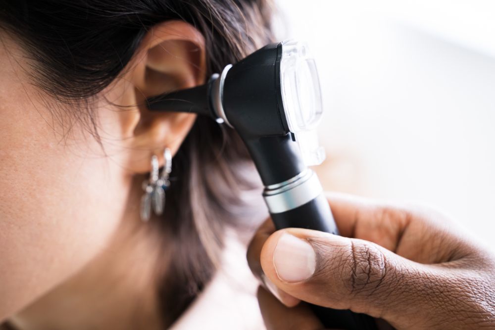
Diagnosis begins with a careful examination of the ear by an ENT or otologist.
Microscopic Ear Examination
A detailed microscopic examination of the ear allows the physician to evaluate the eardrum, ear canal, retraction pockets, drainage, and visible skin debris. This examination may reveal a cholesteatoma or findings that require additional investigation.
Hearing Test
A comprehensive audiogram measures hearing in each ear and determines whether conductive, sensorineural, or mixed hearing loss is present. A hearing test is particularly important when a cholesteatoma has affected the ossicles or other structures involved in sound transmission.
CT Scan of the Temporal Bone
A CT scan of the temporal bone may be used to evaluate the extent of disease and identify erosion or involvement of the mastoid and middle-ear structures. CT imaging can help the surgeon understand the anatomy and plan treatment.
MRI
In selected situations, MRI—particularly specialized diffusion-weighted imaging—may be used to evaluate residual or recurrent cholesteatoma after treatment. The appropriate imaging study depends on the clinical situation.
How Is Cholesteatoma Treated?
Treatment depends on the size and location of the cholesteatoma, whether infection is present, whether hearing has been affected, and whether surrounding structures have been damaged. The primary goal is to remove the cholesteatoma and prevent further damage. Hearing restoration is an important consideration, but it is generally secondary to completely controlling the disease.
Microscopic Ear Cleaning
In selected small or limited cases, an ENT specialist may be able to clean the affected area under microscopic visualization and control associated infection. This does not necessarily eliminate an established cholesteatoma. Continued monitoring is important because cholesteatoma can continue to grow or recur.
Medication
If infection or inflammation is present, medication may be used as supportive treatment. Antibiotics or ear drops may help control an associated infection, but medication does not remove an established cholesteatoma.
Surgery
When a cholesteatoma is established or has progressed beyond what can safely be managed in the office, surgical removal is generally required. Common procedures include:
- Mastoidectomy
- Tympanoplasty
- Ossicular chain reconstruction
The specific operation depends on how far the disease has spread and which structures are involved.
Cholesteatoma Surgery
The purpose of cholesteatoma surgery is to remove the abnormal skin and keratin, control infection, and create a safe, stable ear.
Mastoidectomy
A mastoidectomy removes diseased tissue from the mastoid bone behind the ear. It is commonly performed when a cholesteatoma has extended into the mastoid. Different surgical approaches may be appropriate depending on the extent and location of the disease.
Tympanoplasty
Tympanoplasty repairs the eardrum and, when appropriate, helps reconstruct the middle-ear sound-conduction system. It may be performed together with cholesteatoma removal when the eardrum has been damaged or perforated.
Ossicular Chain Reconstruction
The cholesteatoma may erode one or more of the three tiny middle-ear bones. If the ossicles have been damaged, ossicular chain reconstruction may be performed to rebuild the pathway through which sound travels to the inner ear. Hearing restoration is an important consideration, but the exact reconstruction depends on the condition of the ear at the time of surgery.
Can Cholesteatoma Surgery Restore Hearing?
It can improve hearing in some patients, particularly when conductive hearing loss is caused by damage to the middle-ear sound-conduction system. However, hearing restoration is not guaranteed. The ability to improve hearing depends on factors such as:
- How extensive the cholesteatoma is
- Whether the ossicles are intact
- Whether the eardrum is damaged
- Whether the inner ear has been affected
- The type of reconstruction required
The first objective of surgery is to establish a safe, disease-free ear. Hearing reconstruction may be performed at the same time or considered separately depending on the individual case.
Is Cholesteatoma Surgery Always Necessary?
An established cholesteatoma generally cannot be permanently eliminated with medication alone. Limited disease may sometimes be managed with careful microscopic cleaning and close observation in selected circumstances, but surgical removal is the standard definitive treatment for established cholesteatoma. The appropriate approach depends on the patient’s examination, imaging, symptoms, and extent of disease.
What Happens If Cholesteatoma Is Left Untreated?
A cholesteatoma can continue to enlarge and erode nearby structures. Potential complications include:
- Progressive conductive hearing loss
- Damage to the ossicles
- Persistent ear drainage
- Damage to the inner ear
- Vertigo or balance problems
- Facial nerve injury or facial weakness
- Chronic infection
- Meningitis
- Intracranial infection or abscess
Serious complications are uncommon, but they can occur when disease progresses into surrounding structures. This is why persistent ear drainage or unexplained hearing loss should not simply be treated repeatedly as an ordinary ear infection without determining the underlying cause.
Can a Cholesteatoma Come Back?
Recurrence or residual disease is one reason long-term follow-up is important. Follow-up may include:
- Microscopic ear examinations
- Hearing tests
- Repeat imaging when indicated
- Additional surgery in selected cases
The appropriate surveillance plan depends on the type and extent of the original disease and the surgical approach used.
When Should You See an ENT for Possible Cholesteatoma?
You should consider an ENT evaluation if you experience:
- Persistent or recurrent ear drainage
- Foul-smelling ear discharge
- Repeated ear infections in the same ear
- Unexplained hearing loss
- A persistent feeling of ear pressure or fullness
- Dizziness associated with chronic ear symptoms
- A known eardrum retraction or perforation
- Hearing loss that continues after an ear infection has been treated
Persistent foul-smelling drainage combined with hearing loss deserves particular attention. These symptoms can occur with cholesteatoma and should be evaluated rather than repeatedly treated without identifying the underlying cause.
Why Choose Dr. Neil Sperling for Cholesteatoma Treatment and Surgery
If you have persistent ear drainage, recurrent infections, hearing loss, or a suspected cholesteatoma, a detailed examination can determine whether additional testing or surgical treatment is appropriate.
Dr. Neil Sperling provides evaluation and surgical treatment for hearing and middle-ear disorders, including cholesteatoma, eardrum perforation, conductive hearing loss, and ossicular chain problems. His practice lists cholesteatoma removal among its ear and hearing surgical procedures.
The practice’s current physician information identifies Dr. Sperling as Medical Director of Hearing Healthcare and Technologies, Department of Otolaryngology, Lenox Hill Hospital.
NYC Office Locations
Midtown Manhattan
36A E. 36th Street, Suite 200
New York, NY 10016
Manhattan West Side
9 West 67th Street
New York, NY 10023
Phone: 212-889-8575
Frequently Asked Questions
A cholesteatoma is an abnormal collection of keratinized skin cells and debris trapped in the middle ear, mastoid, or nearby structures. It is not cancerous but can grow and cause damage to surrounding ear structures if untreated.
No, a cholesteatoma is not a malignant tumor or cancer. It is a benign but potentially destructive growth of keratinizing skin tissue that can erode bones and damage ear structures, requiring removal to prevent further harm.
Cholesteatomas commonly develop due to eardrum retraction pockets caused by Eustachian tube dysfunction, chronic ear infections, or a perforated eardrum. Some cases are congenital, meaning they are present from birth without prior ear disease.
Symptoms include persistent or recurrent ear drainage, foul-smelling ear discharge, hearing loss, ear fullness or pressure, ear pain, tinnitus, dizziness, and recurrent ear infections. Some people may have few symptoms until the condition advances.
Yes, cholesteatoma can damage the ossicles—the small bones in the middle ear responsible for sound transmission—leading to conductive hearing loss. Hearing loss severity depends on the extent and location of the cholesteatoma.
Diagnosis involves a detailed microscopic ear examination by an ENT specialist, hearing tests (audiogram), and imaging studies such as CT scans to assess disease extent. MRI may be used in certain cases to detect residual or recurrent cholesteatoma.
Treatment depends on disease size and damage but primarily involves surgical removal to prevent further harm. Small cases may be managed with microscopic cleaning and medication for infection, but surgery is often necessary for established cholesteatomas.
Common surgeries include mastoidectomy to remove diseased mastoid bone tissue, tympanoplasty to repair the eardrum, and ossicular chain reconstruction to restore hearing by repairing damaged middle ear bones. The choice depends on disease extent and structures involved.
Yes, if the cholesteatoma affects inner ear structures involved in balance, it can cause dizziness or vertigo. Persistent dizziness with ear symptoms should be evaluated by an ENT specialist.