
What is Cholesteatoma?
Cholesteatoma is a benign growth caused by trapped skin cells and protein behind the eardrum. When skin from the ear canal penetrates the eardrum, the keratin it produces can accumulate in the middle ear, leading to cholesteatoma. If untreated, it can erode the ossicles, cause hearing loss, and potentially damage the inner ear, leading to more severe complications like facial paralysis or meningitis.
What are the Symptoms?
While some patients may have mild or no symptoms, the most common sign of cholesteatoma is frequent ear infections, often accompanied by a foul-smelling discharge. Hearing loss is also frequently present.
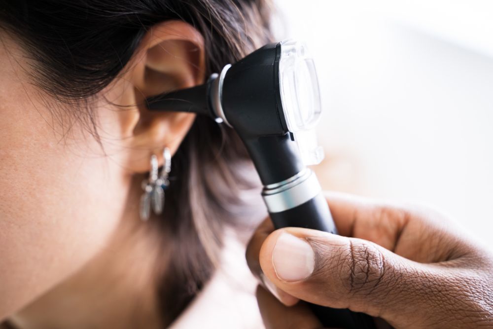
How is it Diagnosed?
Cholesteatoma is diagnosed through a microscopic ear examination during an office visit. A hearing test (audiogram) assesses hearing damage, and a CT scan of the temporal bone checks the extent of cholesteatoma spread.
Treatment and Surgery
Treatment for cholesteatoma focuses on controlling infection and preventing bone damage, with hearing restoration as a secondary goal. Small cases may be cleaned in the office, while advanced cases usually need surgery, including mastoidectomy, tympanoplasty, and ossicular reconstruction. Close follow-up is crucial as cholesteatomas can recur, sometimes requiring additional surgery.
Frequently Asked Questions
Cholesteatoma is a benign growth caused by trapped skin cells and protein behind the eardrum. It occurs when skin from the ear canal penetrates the eardrum, leading to keratin accumulation in the middle ear. If left untreated, it can erode the ossicles, cause hearing loss, and may lead to serious complications like facial paralysis or meningitis.
Many patients experience frequent ear infections accompanied by foul-smelling discharge. Hearing loss is also a common symptom, though some patients may have mild or no symptoms initially. Early diagnosis is important to prevent progression.
Diagnosis involves a microscopic examination of the ear during an office visit to visually assess the eardrum and middle ear. A hearing test (audiogram) is conducted to evaluate hearing loss, and a CT scan of the temporal bone is used to determine the extent of cholesteatoma spread.
Treatment aims to control infection, prevent bone damage, and restore hearing if possible. Small cases may be managed through office cleaning, while advanced cases typically require surgery such as mastoidectomy, tympanoplasty, and ossicular reconstruction. Ongoing follow-up is essential due to risk of recurrence.
Yes, cholesteatoma can erode the ossicles in the middle ear, which are essential for transmitting sound, leading to hearing loss. Early detection and treatment help to minimize the risk of permanent hearing damage.
Cholesteatomas have a tendency to recur even after treatment. Close follow-up appointments and monitoring are crucial to detect and manage any recurrence early, possibly requiring additional surgery to prevent complications.